Resultados
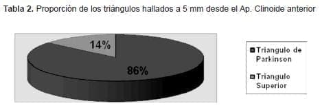
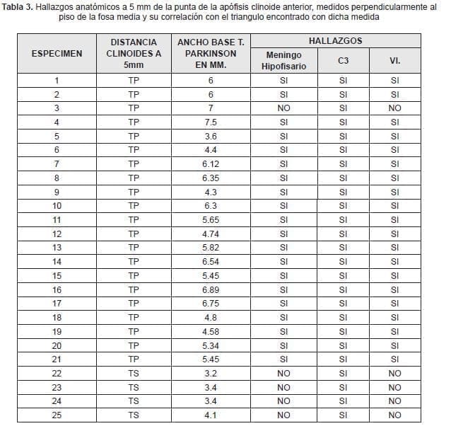
De los 25 especímenes que se disecaron se encontró solo en 4 de ellos el triangulo Superior a 5 mm de la punta del apófisis clinoide anterior medido con una línea recta perpendicular desde el reparo anatómico referido (16%), en el resto de los especímenes se encontró el triangulo de Parkinson (86%) –Tabla 3- (cuyos límites han sido referidos ampliamente en la primera parte), y siempre que se accedió a dicho triangulo se halló :el tronco meningohipofisiario, la porción C3 –horizontal- de la arteria carótida interna intra cavernosa y el sexto par. -Tabla 2-.


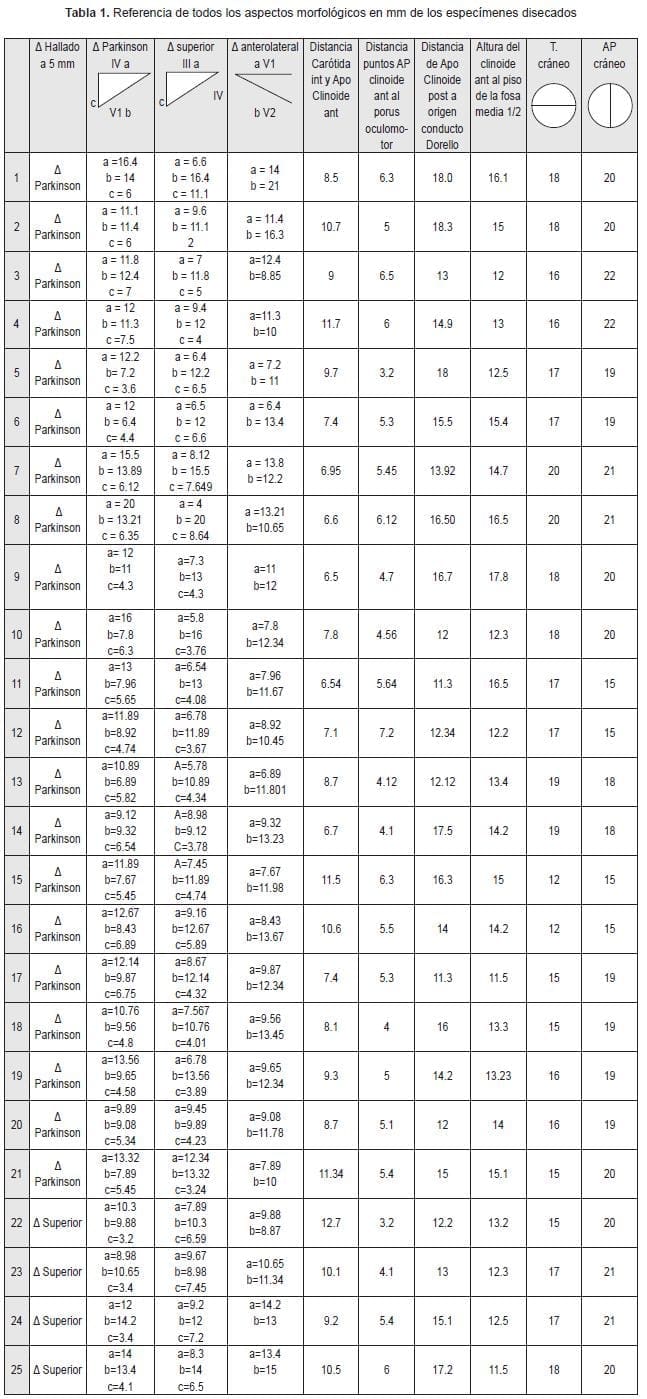
Cuando se encontró el triangulo superior, la arista posterior -ancho del triangulo-, era menor de 4.1 mm, ( promedio de 3.47 mm) Tabla 1 y 2.
En promedio la altura de la fosa media desde la apófisis clinoide anterior fue de 13.89 mm. Tabla 1.
Las medidas promedio del triangulo de Parkinson fueron: (promedios tomados de la Tabla 1) – las medidas de los nervios fueron realizadas tomando como parámetros una línea perpendicular al piso de la fosa media que cae desde la punta del apófisis clinoide anterior, desde dicha línea, se midió la longitud de los pares involucrados hasta su intersección con otros pares formando los triángulos-
Cuarto par (lado a): 12.53 mm
Nervio V 1 (lado b): 10.07 mm
Ancho (lado c): 5.3 mm
Encontramos también en promedio que la distancia desde la cara interna del apófisis clinoide anterior y la cara medial de la emergencia de la arteria carótida interna de subclinoidea a supra clinoidea fue de 8.93 mm (Tabla 1).

Se tomaron también como referencia las medidas en el eje anteroposterior del cráneo y el transverso medido sin el grosor del diploe ( de cara endocraneana a cara endocraneana) como parámetro de comparación. (Tabla 1).
Discusión
El abordaje de lesiones al interior del seno cavernoso siempre ha ofrecido al neurocirujano un grado importante de dificultades, por la cercanía de los pares craneales entre sí, y por el gran numero de estructuras halladas en esta zona anatómica de menos de 2 cm cúbicos. (14, 17, 18, 21, 22, 23, 24, 25, 26 27, 28, 32, 33, 34, 35, 36, 39, 43, 44, 45, 46.). El conocimiento de las distintas estructuras que constituyen la pared del seno permite al neurocirujano utilizarlas como una ventana para acercarse a una estructura en especial y no a otra.
Con esta visión es que hemos hecho nuestro trabajo, el de abrir una ventana que nos ofrezca la posibilidad de entrar en contacto con el mayor numero de estructuras criticas y evitar que dicho abordaje cause más complicaciones que la patología misma.
La ventana a la que nos referimos es el triangulo de Parkinson, (37, 38, 40, 41, 42) el cual fue escogido dentro de las 11 opciones descritas por distintos autores (Fukushima, Dolenc ,etc. “15, 16, 19, 20”) porque es el que en nuestro estudio anatómico nos permitió visualizar la porción transversa de la carótida interna (y por tanto posible vía de acceso para aneurismas saculares de dicho segmento, y aneurismas de hipofisiaria superior inferolaterales,) el tronco meningohipofisiario (aneurismas de dicho segmento) y el sexto par al igual que la cadena simpática (neurofibromas y schwanomas) sin contar que en este triángulo, la duramadre solo tiene una cubierta (y no dos como en los otros triángulos). ( 1, 2, 3, 4, 5, 6, 7, 8, 9, 10, 11, 12, 13).
La duda que nos embargaba era si el espacio de dicho triángulo era optimo para acceder a ese grupo de estructuras intra cavernosas y ,que parámetro podría ser útil usando anatomía macroscópica para acercarse a dicho triangulo, decidimos utilizar el apófisis clinoide anterior y evaluar a través de la altura desde dicho reparo hasta la fosa media con una línea imaginaria que descendiera desde su punta, perpendicular al piso, y encontramos que a 5 mm del apófisis clinoide en esta línea, se halla el triangulo de Parkinson en el 86% de los especímenes y en el 100% de dichos triángulos se encontraron los elementos referidos anteriormente.
Solo en el 14% se encontró el triángulo superior; y cuando a este se llegaba, ninguna de las estructuras referidas eran localizadas.
Hay que tener en cuenta que este estudio no incluyó patologías que deformaran la anatomía habitual del seno, lo que hace suponer que nuestro parámetro podría emplearse cuando la patología a intervenir no modifique mayormente el tamaño habitual del seno, o que sean netamente intra cavernosas, ó cuando las estructuras de la pared del seno se mantienen en la misma posición a pesar de la patología (e.g. algunos meningiomas que nacen de la pared lateral del seno).
Sabemos que para hacer una recomendación quirúrgica se necesitan mas especímenes de disección, pero este trabajo puede ser un buen inicio para la continuación de futuros estudios que tengan en cuenta deformidades a la anatomía normal, o variedades en los diámetros transverso y antero posterior del cráneo que al fin y al cabo son factores que modifican alturas y distancias de porciones mas especificas en la bóveda craneana
Referencias
1. Al mefty O, Smith R. surgery of tumors invading the cavernous sinus. Surg Neurol 30: 370-381, 1988.
2. Al-mefty O, Anand VK: zygomatic approach to skullbase lesions, J Neurosurg 73: 608-673, 1990.
3. Al – mefty O, Borda LA: skull/base/ chordomas: a management/challenge. J Neurosurg 86: 182- 189. 1997.
4. Ammirati, Bernando A: analytical evaluation of complex anterior approaches to the cranial base: An anatomic study. Neurosurg 43: 1398-1408, 1998.
5. Ammirati M, Ma J, Cheatham ML, et al: The mandibular swing – Transcervical approach to the skull base : anatomical study. Technical note. J Neurosurg 78: 673 – 681,1993.
6. Barrow DL , Spector RH, Braun IF, et al. Classification and treatment of spontaneous carotid-cavernous sinus fistulas. J Neurosurg 62: 248-256, 1985.
7. Bouchet A.- Cuilleret J.: Cráneo Óseo, Conducto Raquídeo. Anatomía descriptiva, Topográfica y funcional Tomo Sistema Nervioso Central :1:7-40 7:156-171,1997.
8. Chang SD, Steinberg GK superficial tempor al artery to middle cerebral artery anastomosis Tech Neurosurg 6:86-100, 2000.
9. Day al: aneurysms of the ophthalmic segment J. neurosurg 72: 677-691, 1990.
10. Day JD, Fukushima T, Tiamotta Sc: microanatomical study of the extradural middle fossa approach to the petroclival and posterior cavernous sinus region: description of the rhomboid constructs Neurosurgery 34: 1009 – 1016, 1994.
11. Debrun GM, Vinuela F, Fox AJ et al. Indications for treatment and classifications of 132 carotid-cavernous fistulas Neurosurgery. 22: 285-289, 1988.
12. Demorais Jy, Lana-Peixotoma Bilateral Intracavernous carotid aneurysms treatment by bilateral carotid ligation Surg Neurol 1978.
13. Diaz Day J. Apuzzo J. L. Michael Koos Wolfgang The transoral approach microsurgical dissection of the cranial base the craniocervical junction and foramen magnun chapter 5; 124-134, 1996.
14. Diaz FG, Ohaegbulam S, Dujovny M, et al. Surgical alternatives in the treatment of cavernous sinus aneurysms J Neurosurg 71: 846-853, 1989.
15. Dolenc V. Direct microsurgical repair of intracavernous vascular lesions. J Neurosurg. 58: 824 – 831, 1983.
16. Dolenc V. cavernous sinus masses. In Apuzzo ML (ed) Brain surgery: complication avoidance and management Churchill Livingstone PP 60/ 614, 1993.
17. Francis PM, Zabramski JM, Spetzler RF, et al. treatment of carotid-cavernous fistulas: part II surgical interventions BNI. Q. 7: 7-15, 1991.
18. Franco DeMonte, Díaz Eduardo, Callender David, Suk Ian.: Transmandibular, circumglossal, re-tropharyngeal approach for chordomas of the clivus and upper cervical spine. Neurosurg Focus 10 (3): Vol.10, March 2001: 1-5.
19. Fukushima T. Direct operative approach to the vascular lesions in the cavernous sinus: Summary of 27 cases Mt fusi workshop cerebrovas Dis G; 169- 189, 1988.
20. Fukushima T, day JD Tung H intracavernous carotid artery aneurysms in Apuzzo (Ed) Brain surgery: complication avoidance and management Churchill Livingstone Inc New York PP 925-944, 1992.
21. Glasscock ME: Exposure of the intra- petrous portion of the carotid artery. In Hamburger CA wersall (Eds): Disorders of the skull base region: proceedings of thel0th Nobel Symposium, Stockholm, 1968. Stockholm almgvist and wicfsell, pp13f143, 1969.
22. Hamby WB. carotid – cavernous fistula. Report of 32 surgically treated cases and suggestions for definitive operation. J Neurosurg. 21: 859-866, 1964.
23. Hirsch wcjr hryshko FG sekhar LN, Brunberg J. Comparison of MR imaging. Ct and angiography in the evaluation of the enlarged cavernous sinus: a microsurgical study. Neurosurgery 26: 903- 932, 1990.
24. Hitotsumatsu T, Rhoton AL Jr: Unilateral upper and lower subtotal maxillectomy approaches to the skull base: Microsurgical Anatomy. Neurosurgery 46:1416- 1453, 2000.
25. Hitotsumatsu T, Rhoton AL Jr,Matsushima T: Surgical Anatomy of the midface and the midline skull base, in Spetzler RF (Ed) Opperative Techniques in Neurosurgery. 1999, Vol. 2, pp 160 – 180.
26. Kawakami K. Yamanouchi Y, Kawamura Y, Matsumura H: operative approach to the frontal skull base: extensive transbasal approach neurosurgery 28: 720-725, 1991.
27. Kawase T, Toya S, Shiobara R, et al: Transpetrosal approach for aneurysms of the lower basilar artery Neurosurg 63: 857 – 861, 1985.
28. Kawase T, Shiobara R, Toya S: anterior transpetrosal transtentorial approach for spheno petroclival meningiomas: surgical method and results in 10 patients Neurosurgery 28: 869-876. 1991.
29. Latarjet – Ruiz Liard: Huesos del Cráneo. Anatomía Humana Tomo 1: III: 69-88,1992.
30. Lawton MT, Hamilton MG Morcos JJ, et al Revascularization and aneurysm surgery current techniques, indications, and outcomes. Neurosurgery 38: 83-94 1996.
31. Linskey ME, Sekhar LN. Cavernous sinus hemangiomas a series: a series, a review, and a hypothesis neurosurgery 30: 101-107, 1992.
32. Linskey ME, Sekhar LN, Hirsch Jr, WL et al aneurysms of the intracavernous carotid artery: clinical presentation, radiographic features, and pathogenesis. Neurosurgery. 26: 71-79, 1990.
33. Linskey ME, Sekhar LN. Horton JA, et al. Aneurysms of the intracavernous carotid artery: a multidisciplinary approach to treatment J. Neurosurg 75: 525- 534, 1991.
34. Linskey ME, Sekhar LN, Hirsch Jr, WL et al aneurysms of the intracavernous carotid artery: natural history and indications for treatment. Neurosurgery 26: 933-938, 1990.
35. Lombardi D, Giovanelli M, de tribolet N Sellar and parasellar extra axial cavernous hemangiomas. Acta neurochir (Wien) 130, 47-54: 1994.
36. Oliveira E, Rhoton AL Jr,Peace DA:Microsurgical Anatomy of the region of the foramen magnum. Surg Neurol 24: 293- 352,1985.
37. Parkinson D. A surgical approach to the cavernous portions of the carotid artery. Anatomical studies and case report. J neurosurg.; 23: 474- 483.1965.
38. Parkinson D. carotid cavernous fistula: direct repair with preservation of the carotid artery. Technical note. J neurosurg; 38: 99 – 106, 1973.
39. Poppen : Head. An Atlas of Neurosurgical Techniques Part I. Section 4:292 – 303,1960.
40. Rhoton Albert L.: The Foramen Magnum. Neurosurgery The posterior cranial Fossa: Microsurgical anatomy & surgical Approaches, Vol. 47, No.3, September 2000 Supplement: S155 – S193.
41. Rhoton AL Jr., Buza R: Microsurgical Anatomy of the jugular foramen. J Neurosurg 42: 541 – 550, 1975.
42. Rhoton AL the cavernous sinus the cavernous venous plexus and the carotid collar neurosurgery 5 (suppl): 375- c10 2002.
43. Roski RA Spetzler RF Nulsen FE late complications of carotid ligation in the treatment of intracranial aneurysms J. neurosurg 54: 583-587. 1981.
44. Seckhar LN, Nanda A, Sen CN, Snyderman CN, Janecka IP: The extended frontal approach to tumors of the anterior, middle and posterior skull base. J Neurosurg 76: 198 – 206, 1992.
45. Spetzler RF, Fukushima T, martin N et al petrous carotid to- intradural carotid saphenous vein graft for intracavernous Sgiant aneurysm tumor, and occlusive cerebrovascular disease. J Neurosurg 73: 496-501. 1990.
46. Zabramski JM, Kiris T Sankla SK et al. Orbizygomatic craniotomy technical note neurosurg 62: 667-672. 1985.

