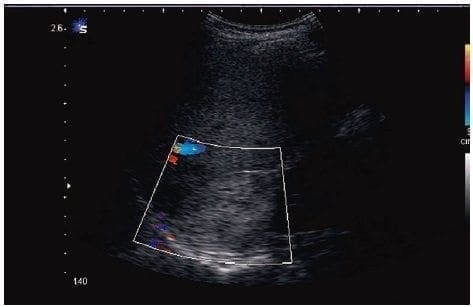
Diversos procesos renales pueden ser detectados mediante la ecografía, la cual podría ser útil en la práctica quirúrgica, sobre todo en la aproximación diagnóstica.
La hidronefrosis puede ser obstructiva o no obstructiva. La urolitiasis es la causa más común
de las obstructivas y produce focos de ecogenicidad bien definida con sombra acústica posterior y un artefacto de centelleo posterior con el ultrasonido Doppler.
La enfermedad quística renal puede ser de origen hereditario o no hereditario. El diagnóstico diferencial puede aclararse con base en la presencia de nefromegalia, de quistes discretos o de numerosos microquistes, y la demostración de compromiso multiorgánico.
Las masas renales que no son tumorales ni quísticas (abscesos, hematomas) pueden detectarse ecográficamente. Sin embargo, la mayor utilidad de la ecografía renal radica en la identificación de neoplasias renales, lo cual es particularmente útil para decidir la intervención quirúrgica urológica. A pesar de que la tomografía computarizada y la resonancia magnética son frecuentemente usadas para caracterizar las lesiones, la ecografía con contraste está emergiendo como una modalidad de imagen alternativa.
Cumplimiento de normas éticas
Consentimiento informado:
Este estudio es una revisión de la literatura, y como tal no hay necesidad de un consentimiento informado ni de aprobación del Comité de Ética Institucional.
Conflictos de interés: Ninguno reportado por los autores.
Fuentes de financiación: autofinanciado por los autores.
Referencias
- 1. Sigel MJ. Operative ultrasound in general surgery. Am J Surg. 1996;172:15-20.
- 2. Prada R. Historia del diagnóstico por ultrasonido. Revista de la Facultad de Medicina de la Universidad Nacional de Colombia. 1995;43:204-6.
- 3. Newman PG, Rozycki GS. The history of ultrasound. Surg Clin North Am. 1998;78:179-95.
- 4. White DN. Neurosonology pioneers. Ultrasound Med Biol. 1988;14:541-61.
- 5. Wells PN. Developments in medical ultrasonics. World Med Electron. 1966;4:2721.
- 6. Wild JJ. The use of ultrasonic pulses for the measurement of biologic tissues and the detection of tissue density changes. Surgery. 1950;27:183-7.
- 7. Wild JJ, Reid JM. Diagnostic use of ultrasound. Br J Phys Med. 1956;19:248-57.
- 8. Wild JJ, Reid JM. Further pilot echographic studies of the histologic structure of tumors of the living intact human breast. Am J Pathol. 1952;28:839.
- 9. Eiseman B, Greenlaw RH, Gallagher JQ. Localization of common duct stones by ultrasound. Arch Surg. 1965;91:195-5. doi:10.1001/archsurg.1965.01320130197023.
- 10. Makuuchi M, Hasagawa H, Yamasaki S. Newly devised intra-operative probe. Image technology and information display. Medical. 1979;11:1167-8.
- 11. Machi J, Sigel B. Intraoperative ultrasonography. Radiol Clin North Am. 1992;30:1085-1103.
- 12. Machi J, Sigel B, Zaren HA, Kurohiji T, Yamashita Y. Operative ultrasonography during hepatobiliary and pancreatic surgery. World J Surg. 1993;17:640-6.
- 13. Schlegel JU, Diggdon P, Cuéllar J. The use of ultrasound for localizing renal calculi. J Ural. 1961;86:367-9.
- 14. Sigel B, Kraft AR, Nyhus LM, Coelho JC, Gavin MP, Spigos DG. Identification of a parathyroid adenoma by operative ultrasonography. Arch Surg. 1981;116:234-5.
- 15. Tanaka K, Ito K, Wagai T. The localization of brain tumors by ultrasonic techniques. A clinical review of 111 cases. J Neurosurg. 1965;23:135-47.
- 16. Whitsett MC. Ultrasound imaging and advances in system features. Ultrasound Clin. 2009;4:391-401.
- 17. Beggs AD, Thomas PR. Point of use ultrasound by general surgeons: Review of the literature and suggestions for future practice. Int J Surg. 2013;11:12-7. doi: 10.1016/j. ijsu.2012.11.014.
- 18. Freitas ML, Frangos SG, Frankel HL. The status of ultrasonography training and use in general surgery residency programs. J Am Coll Surg. 2006;202:453-8.
- 19. Nassour I, Spalding MC, Hynan LS, Gardner AK, Williams BH. The surgeon-performed ultrasound: A curriculum to improve residents basic ultrasound knowledge. J Surg Res. 2017;213:51-9. doi: 10.1016/j. jss.2017.02.031.
- 20. Williams RJ, Windsor AC, Rosin RD, Mann DV, Crofton M. Ultrasound scanning of the acute abdomen by surgeons in training. Ann R Coll Surg Engl. 1994;76:228e-33.
- 21. Asociación Colombiana de Radiología. Historia de la radiología. Fecha de consulta: 29 de julio de 2019. Disponible en: https://www.acronline.org/Nosotros/Historia-de-la-Radiolog%C3%ADa
- 22. Granada JC. Concordancia del diagnóstico ecográfico de patología biliar entre el residente de cirugía general y el radiólogo en el Hospital Simón Bolívar durante 1998 a 1999 (tesis de). Santa Fe de Bogotá: Universidad El Bosque; 2000.
- 23. Salcedo Y, Segura A, Rodríguez A, Segura JM. Anatomía ecográfica abdominal normal. Sistemática de exploración. Semergen. 2014;40:205-10.
- 24. Lindelius A, Törngren S, Sondén A, Pettersson H, Adami J. Impact of surgeon-performed ultrasound on diagnosis of abdominal pain. Emerg Med J. 2008;25:486-91.
- 25. Lindelius A, Törngren S, Pettersson H, Adami J. Role of surgeon-performed ultrasound on further management of patients with acute abdominal pain: A randomised controlled clinical trial. Emerg Med J. 2009;26:561-6.
- 26. Lindelius A, Törngren S, Pettersson H, Adami J. Patient factors influencing the effect of surgeon-performed ultrasound on the acute abdomen. Crit Ultrasound J. 2010;2:97-105.
- 27. Di Lelio A, Cestari C, Lomazzi A, Beretta L. Cirrhosis: Diagnosis with sonographic study of the liver surface. Radiology. 1989;172:389-92.
- 28. Freeman MP, Vick CW, Taylor KJ, Carithers RL, Brewer WH. Regenerating nodules in cirrhosis: Sonographic appearance with anatomic correlation. Am J Roentgenol. 1986;146:533-6.
- 29. Simonovsky V. The diagnosis of cirrhosis by high resolution ultrasound of the liver surface. Br J Radiol. 1999;72:29-34.
- 30. Harbin WP, Robert NJ, Ferrucci JT Jr. Diagnosis of cirrhosis based on regional changes in hepatic morphology: A radiological and pathological analysis. Radiology. 1980;135:273-83.
- 31. Giorgio A, Amoroso P, Lettieri G, Fico P, de Stefano G, Finelli L. Cirrhosis: Value of caudate to right lobe ratio in diagnosis with US. Radiology. 1986;161:443–5. doi: 10.1148/radiology.161.2.3532188
- 32. Lim JH. Dysplastic nodules in liver cirrhosis: Detection with triple phase helical dynamic CT. Br J Radiol. 2004;77:911-6.
- 33. Lim JH, Choi BI. Dysplastic nodules in liver cirrhosis: Imaging. Abdom Imaging. 2002;27:117-28.
- 34. Segura A, Valero I, Díaz N, Segura JM. Ecografía hepática: lesiones focales y enfermedades difusas. Semergen. 2016;42:307-14.
- 35. Kim TK, Jang H-J, Wilson SR. Hepatic neoplasms: Features on grayscale and contrast enhanced ultrasound. Ultrasound Clin. 2007;2:333-54.
- 36. Tanaka S, Kitamura T, Imaoka S, Sasaki Y, Taniguchi H, Ishiguro S. Hepatocellular carcinoma: Sonographic and histologic correlation. Am J Roentgenol. 1983;140:701-7.
- 37. Yoshikawa J, Matsui O, Takashima T, Ida M, Takanaka T, Kawamura I, et al. Fatty metamorphosis in hepatocellular carcinoma: Radiologic features in 10 cases. Am J Roentgenol. 1988;151:717-20.
- 38. Vachha B, Sun MR, Siewert B, Eisenberg RL. Cystic lesions of the liver. Am J Roentgenol. 2011;196:W355-66.
- 39. Colli A, Cocciolo M, Mumoli N, Cesarini L, Prisco A, Gaffuri I, et al. Peripheral intrahepatic cholangiocarcinoma: Ultrasound findings and differential diagnosis from hepatocellular carcinoma. Eur J Ultrasound. 1998;7:93-9.
- 40. Chung YE, Kim MJ, Park YN, Choi JY, Pyo JY, Kim YC, et al. Varying appearances of cholangiocarcinoma: Radiologic-pathologic correlation. Radiographics. 2009;29:683-700.
- 41. Robledo R, Muro A, Prieto ML. Extrahepatic bile duct carcinoma: US characteristics and accuracy in demonstration of tumors. Radiology. 1996;198:869-73.
- 42. Lee WJ, Lim HK, Jang KM, Kim SH, Lee SJ, Lim JH, et al. Radiologic spectrum of cholangiocarcinoma: Emphasis on unusual manifestations and differential diagnoses. Radiographics. 2001;21(Spec number):S97-116.
- 43. Jhaveri KS, Halankar J, Aguirre D, Haider M, Lockwood G, Guindi M, et al. Intrahepatic bile duct dilatation due to liver metastases from colorectal carcinoma. Am J Roentgenol. 2009;193:752-6.
- 44. Lev-Toaff AS, Bach AM, Wechsler RJ, Hilpert PL, Gatalica Z, Rubin R. The radiologic and pathologic spectrum of biliary hamartomas. Am J Roentgenol. 1995;165:309-13.
- 45. Zheng RQ, Kudo M, Onda H, Inoue T, Maekawa K, Minami Y, et al. Imaging findings of biliary hamartomas (von Meyenburg complexes). J Med Ultrason. 2005;32:205-12.
- 46. Avlonitis VS, Linos D. Primary hepatic lymphoma: A review. Eur J Surg. 1999;165:725-9.
- 47. Nghiem HV, Bogost GA, Ryan JA, Lund P, Freeny PC, Rice KM. Cavernous hemangiomas of the liver: enlargement over time. Am J Roentgenol. 1997;169:137-40.
- 48. Benter T, Kluhs L, Teichgraber U. Sonography of the spleen. J Ultrasound Med. 2011;30:1281-93.
- 49. Spielmann AL, DeLong DM, Kliewer MA. Sonographic evaluation of spleen size in tall healthy athletes. Am J Roentgenol. 2005;184:45-9.
- 50. Sutherland T, Temple F, Hennessy O, Lee WK. Abdomen’s forgotten organ: Sonography and CT of focal splenic lesions. J Med Imaging Radiat Oncol. 2010;54:120-8.
- 51. Saad NEA, Saad WEA, Davies MG, Waldman DL, Fultz PJ, Rubens DJ. Pseudoaneurysms and the role of minimally invasive techniques in their management. Radiographics .2005;25(Suppl.1):S173-89.
- 52. Weingarten MJ, Fakhry J, McCarthy J, Freeman SJ, Bisker JS. Sonography after splenic embolization: The wedge-shaped acute infarct. Am J Roentgenol. 1984;142:957-9.
- 53. Goerg C, Schwerk WB. Splenic infarction: Sonographic patterns, diagnosis, follow-up, and complications. Radiology. 1990;174:803-7.
- 54. Giovagnoni A, Giorgi C, Goteri G. Tumours of the spleen. Cancer Imaging. 2005;5:73-7.
- 55. Willcox TM, Speer RW, Schlinkert RT, Sarr MG. Hemangioma of the spleen: Presentation, diagnosis, and management. J Gastrointest Surg. 2000;4:611-3.
- 56. Good LI, Edell SL, Soloway RD, Trotman BW, Mulhern C, Arger PA. Ultrasonic properties of gallstones. Effect of stone size and composition. Gastroenterology. 1979;77:258-63.
- 57. Ralls PW, Colletti PM, Lapin SA, Chandrasoma P, Boswell WD Jr, Ngo C, et al. Real-time sonography in suspected acute cholecystitis. Prospective evaluation of primary and secondary signs. Radiology. 1985;155:767-71.
- 58. Simeone JF, Brink JA, Mueller PR, Compton C, Hahn PF, Saini S, et al. The sonographic diagnosis of acute gangrenous cholecystitis: Importance of the Murphy sign. Am J Roentgenol. 1989;152:289-90.
- 59. Bree RL. Further observations on the usefulness of the sonographic Murphy sign in the evaluation of suspected acute cholecystitis. J Clin Ultrasound. 1995;23:169-72.
- 60. Shea JA, Berlin JA, Escarce JJ, Clarke JR, Kinosian BP, Cabana MD, et al. Revised estimates of diagnostic test sensitivity and specificity in suspected biliary tract disease. Arch Intern Med. 1994;154:2573-81.
- 61. Stefanidis D, Sirinek KR, Bingener J. Gallbladder perforation: Risk factors and outcome. J Surg Res. 2006;131:204-8.
- 62. Teefey SA, Baron RL, Bigler SA. Sonography of the gallbladder: Significance of striated (layered) thickening of the gallbladder wall. Am J Roentgenol. 1991;156:945-7.
- 63. Teefey SA, Dahiya N, Middleton WD, Bajaj S, Dahiya N, Ylagan L, et al. Acute cholecystitis: Do sonographic findings and WBC count predict gangrenous changes? Am J Roentgenol. 2013;200:363-9.
- 64. Rodríguez C, Aldana G. El síndrome de compresión biliar extrínseca benigna o síndrome de Mirizzi: experiencia de cinco años en el Hospital de San José. Rev Colomb Cir. 2008;23:6-11.
- 65. Nakeeb A, Pitt HA, Sohn TA, Coleman J, Abrams RA, Piantadosi S. Cholangiocarcinoma. A spectrum of intrahepatic, perihilar, and distal tumors. Ann Surg. 1996;224:463-75.
- 66. Wilson SR, Novak KL. Sonography of the bowel. Ultrasound Clin. 2014;9:751-73.
- 67. Marco SF, Fernández P, Gil S. La ecografía del tracto gastrointestinal en los pacientes adultos. Med Integr. 2000;35:424-32.
- 68. Atri M, Finnegan PW. The pancreas. In: Rumack CM, Wilson SR, Charboneau JW, editors. Diagnostic ultrasound. Third edition. St. Louis (MO): Mosby; 2005. p. 213-67.
- 69. Nichols MT, Russ PD, Chen YK. Pancreatic imaging: Current and emerging technologies. Pancreas. 2006;33:211-20.
- 70. Martínez-Noguera A, D’Onofrio M. Ultrasonography of the pancreas. 1. Conventional imaging. Abdom Imaging. 2006;32:136-49.
- 71. Lane BF, Wong-You-Cheong JJ. Sonography of the retroperitoneum. Ultrasound Clin. 2014;9:13-7.
Fecha de recibido: 16/10/2018 – Fecha aceptación: 7/05/2019
Correspondencia: Laura Cristancho, Calle 34 N° 98-B-35, torre 7, apartamento 504, Santiago de Cali, Colombia, Teléfonos: (315)
403-2381. Correo electrónico: laura_abc73@hotmail.com
Citar como: Cristancho L, Granada JC. Ecografía en cirugía general. Rev Colomb Cir. 2019;34:372-85. https://doi.org/10.30944/20117582.517.
Este es un artículo de acceso abierto bajo una Licencia Creative Commons – BY-NC-ND https://creativecommons.org/licenses/by-nc-nd/4.0/deed.es