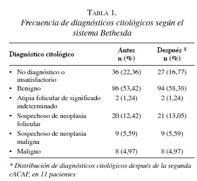
Los resultados de la aCAF fueron reclasificados como: benigno, 103 (63,98 %), maligno, 8 (4,97 %), sospechoso, 32 (19,87 %), e indeterminado, 18 (11,18 %).
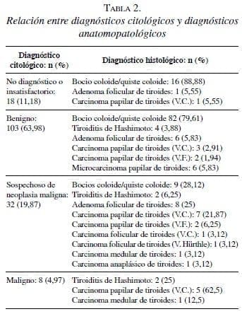
En 136 (84,47 %) pacientes, la enfermedad fue benigna, mientras que en 25 (15,53 %) el resultado fue de carácter maligno (tabla 2). De estos 136 reportes histopatológicos benignos, en 17 (12,5 %) casos la citología fue insatisfactoria, en 98 (72,06 %), fue benigna, en 19 (13,97 %), fue sospechosa de neoplasia maligna, y en 2 (1,47%), fue maligna. De los 25 casos reportados como malignos, 5 (20 %) habían sido reportados como benignos por la citología, 13 (52 %), como sospechosos de neoplasia maligna, 1 (4 %), como indeterminado, y 6 (24 %), como malignos. La distribución de los diagnósticos histopatológicos se muestra en la tabla 2.
Basados en los resultados anteriores (tabla 3), se calcularon los siguientes parámetros, excluyendo e incluyendo los microcarcinomas, respectivamente: sensibilidad, 54,5 % (6/11) y 35,2 % (6/17); especificidad, 98 % (98/100) y 97,8 % (92/94); falso positivo, 1,8 % (2/111) para ambos casos; falso negativo, 4,5 % (5/111) y 9,9 % (11/111); valor diagnóstico positivo, 75 % (6/8) para ambos casos; valor diagnóstico negativo, 98 % (98/100) y 97,8 % (92/94); y precisión diagnóstica global (overall value), 93 % (104/111) y 88,2 % (98/111). El índice kappa para concordancia diagnóstica, fue de 0,598 y 0,423.

Discusión
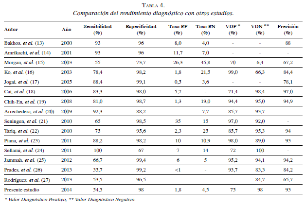
La citología obtenida mediante aspiración con aguja fina posee una sensibilidad que varía en un rango de entre el 55 % y el 100 %, y una especificidad que varía entre el 74 % y el 98 % 13-27 (tabla 4).
En este estudio, al incluir los microcarcinomas papilares, la sensibilidad de la citología obtenida mediante aspiración con aguja fina, fue del 35,2 %, un valor significativamente más bajo que el reportado en la literatura científica. Por otro lado, se encontraron: especificidad 97,8 %, valor diagnóstico positivo de 75 %, valor diagnóstico negativo de 97,8 % y precisión diagnóstica de 88,2 %, valores similares a los rangos reportados en otros estudios 9,28,29. El número de falsos negativos fue considerable con un total de 11 casos, incluyendo microcarcinomas, y de 5, excluyéndolos. Hubo dos falsos positivos, en los cuales se practicó una intervención radical innecesaria. La baja sensibilidad encontrada en este estudio puede explicarse por las siguientes razones: la citología obtenida mediante aspiración con aguja fina es una prueba diagnóstica que depende del operador y, en nuestra institución, sólo a partir del 2010 se practicaron con guía ecográfica, como se recomienda en la literatura científica 30. Lo anterior se suma a la naturaleza de centro de práctica y formación de especialidades médico-quirúrgicas, como radiología y cirugía general, lo cual puede ser uno de los factores que afectó los resultados del presente estudio.
Por otro lado, las variaciones de las características anatómicas de las lesiones son determinantes al momento de obtener una muestra citológica satisfactoria, especialmente,
al discriminar lesiones nodulares menores de 10 mm (microcarcinomas papilares) en el contexto de bocios multinodulares y procesos autoinmunitarios de la glándula tiroides 4. Además, 12 pacientes con diagnóstico citológico insatisfactorio fueron intervenidos quirúrgicamente, lo cual puede explicarse porque los síndromes compresivos y la solicitud de una intervención quirúrgica por parte del paciente, ya sea por motivos estéticos o por el temor a una lesión maligna, son otras indicaciones para que el cirujano practique el procedimiento. Sin embargo, dado al carácter retrospectivo de este estudio, dichas variables no se evaluaron. El alto porcentaje de pacientes con diagnósticos insatisfactorios podría explicar la baja sensibilidad encontrada. Es posible que, al disminuir la incidencia de esta categoría diagnóstica, aumente la sensibilidad de la prueba 31 .
Conclusión
La citología obtenida mediante aspiración con aguja fina es una herramienta con gran especificidad para el diagnóstico de las lesiones de la glándula tiroides. Su baja sensibilidad se debe a la variabilidad de las características anatómicas de las lesiones y a que es una prueba que depende del operador. Una alternativa para aumentar la sensibilidad, es disminuir la frecuencia de diagnósticos indeterminados. El índice kappa muestra una fuerza de concordancia moderada, cuando se excluyen los microcarcinomas (0,598), y débil, cuando se incluyen (0,423).
Diagnostic Performance of Fine Needle Aspiration in Patients with Single Thyroid Nodule at Hospital Universitario del Caribe, Cartagena, Colombia
Abstract
Background: Fine needle aspiration cytology is the best diagnostic tool to choose the conduct regarding a thyroid nodule. Determination of the diagnostic performance of this procedure supports this fact and helps to recognize the behavior of this pathology in our environment.
Methods: A retrospective review of medical records of patients with the diagnostic impression of thyroid nodule in the period of 2007 – 2013 that had fine needle aspiration cytology and were managed surgically was conducted. Sensibility, specificity, positive predictive value, negative predictive value, diagnostic accuracy and concordance were determined in these patients.
Results: The study population was conformed by 161 patients. The following parameters were determined: sensibility, 54,5%; specificity, 98%; false positive rate, 1.8%; false negative rate, 4.5%; positive predictive value, 75%; negative predictive value, 98%; diagnostic accuracy, 93%; and Kappa index, 0.598 excluding micro carcinomas.
Conclusion: Fine needle aspiration cytology is a diagnostic test with high specificity for the diagnosis of thyroid gland nodular lesions. Nonetheless, the variability of the anatomic lesions and the fact that it is an operator dependent test decreases its sensibility, and therefore the histopathologic diagnosis is the gold standard for the definitive diagnosis of thyroid gland lesions.
Key words: Thyroid gland; thyroid nodule; biopsy, fine-needle; cytology; diagnosis; sensitivity and specificity.
Referencias
1. American Thyroid Association Guidelines Taskforce on Thyroid Nodules and Differentiated Thyroid Cancer, Cooper DS, Doherty GM, Haugen BR, Kloos RT, Lee SL, et al. Revised American Thyroid Association management guidelines for patients with
thyroid nodules and differentiated thyroid cancer. Thyroid. 2009;19:1167-214.
2. Román-González A, Restrepo-Giraldo L, Alzate-Monsalve C, Vélez A, Gutiérrez-Restrepo J. Nódulo tiroideo, enfoque y manejo. Revisión de la literatura. Iatreia. 2013;26:197-206. 3. Hamberger B, Gharib H, Melton LJ, 3rd, Goellner JR, Zinsmeister
AR. Fine-needle aspiration biopsy of thyroid nodules. Impact on thyroid practice and cost of care. Am J Med. 1982;73:381-4.
4. Jo VY, Stelow EB, Dustin SM, Hanley KZ. Malignancy risk for fine-needle aspiration of thyroid lesions according to the Bethesda System for Reporting Thyroid Cytopathology. Am J Clin Pathol. 2010;134:450-6.
5. Cibas ES. Fine-needle aspiration in the work-up of thyroid nodules. Otolaryngol Clin North Am. 2010;43:257-71.
6. Pedroza A. Manejo del nódulo tiroideo: revisión de la literatura. Rev Colomb Cir. 2008;23:100-11.
7. Gharib H, Goellner JR. Fine-needle aspiration biopsy of thyroid nodules. Endocr Pract. 1995;1:410-7.
8. Gharib H, Goellner JR, Johnson DA. Fine-needle aspiration cytology of the thyroid. A 12-year experience with 11,000 biopsies. Clin Lab Med. 1993;13:699-709.
9. Gharib H, Goellner JR. Fine-needle aspiration biopsy of the thyroid: An appraisal. Ann Intern Med. 1993;118:282-9.
10. Danese D, Sciacchitano S, Farsetti A, Andreoli M, Pontecorvi A. Diagnostic accuracy of conventional versus sonographyguided fine-needle aspiration biopsy of thyroid nodules. Thyroid. 1998;8:15-21.
11. Cibas ES, Ali SZ, Conference NCITFSotS. The Bethesda System for Reporting Thyroid Cytopathology. Am J Clin Pathol. 2009;132:658-65.
12. Cooper DS, Doherty GM, Haugen BR, Kloos RT, Lee SL, Mandel SJ, et al. Management guidelines for patients with thyroid nodules and differentiated thyroid cancer. Thyroid. 2006;16:109-42.
13. Bakhos R, Selvaggi SM, DeJong S, Gordon DL, Pitale SU, Herrmann M, et al. Fine-needle aspiration of the thyroid: Rate andcauses of cytohistopathologic discordance. Diagn Cytopathol. 2000;23:233-7.
14. Amrikachi M, Ramzy I, Rubenfeld S, Wheeler TM. Accuracy of fine-needle aspiration of thyroid. Arch Pathol Lab Med. 2001;125:484-8.
15. Morgan JL, Serpell JW, Cheng MS. Fine-needle aspiration cytology of thyroid nodules: How useful is it? ANZ J Surg. 2003;73:480-3.
16. Ko HM, Jhu IK, Yang SH, Lee JH, Nam JH, Juhng SW, et al. Clinicopathologic analysis of fine needle aspiration cytology of the thyroid. A review of 1,613 cases and correlation with histopathologic diagnoses. Acta Cytol. 2003;47:727-32.
17. Jogai S, Al-Jassar A, Temmim L, Dey P, Adesina AO, Amanguno HG. Fine needle aspiration cytology of the thyroid: A cytohistologic study with evaluation of discordant cases. Acta Cytol. 2005;49:483-8.
18. Cai XJ, Valiyaparambath N, Nixon P, Waghorn A, Giles T, Helliwell T. Ultrasound-guided fine needle aspiration cytology in the diagnosis and management of thyroid nodules. Cytopathology. 2006;17:251-6.
19. Chih-En T, Chang-Kuo W, Chin-Sung K, Shih-Tang Y, Pin-Fan C, Wei-Cheng L, et al. Fine needle aspiration cytology of thyroid nodules: Evaluation of diagnostic accuracy. Tzu Chi Med J. 2008;20:296-303.
20. Arrechedera-Pacheco C, Oviedo N, Linares Y, Sanabria E, Canozo L. La punción con aguja fina método de estudio diagnóstico en nódulos de la glándula tiroides. Rev Venez Oncol. 2009;21:77-84.
21. Seningen JL, Nassar A, Henry MR. Correlation of thyroid nodule fine-needle aspiration cytology with corresponding histology at Mayo Clinic, 2001-2007: An institutional experience of 1,945 cases. Diagn Cytopathol. 2012;40(Suppl.1):E27-32.
22. Tariq M, Zafar-Iqbal M, Zulifqar-Ali M, Anwar-Ch M, Sulman-Khan R, Irum S. FNAC of thyroid nodule; Diagnostic accuracy of fine needle aspiration cytology (FNAC). Professional Med J. 2010;17:589-97.
23. Piana S, Frasoldati A, Ferrari M, Valcavi R, Froio E, Barbieri V, et al. Is a five-category reporting scheme for thyroid fine needle aspiration cytology accurate? Experience of over 18,000 FNAs reported at the same institution during 1998-2007. Cytopathology.
2011;22:164-73.
24. Sellami M, Tababi S, Mamy J, Zainine R, Charfi A, Beltaief N, et al. Interest of fine-needle aspiration cytology in thyroid nodule. Eur Ann Otorhinolaryngol Head Neck Dis. 2011;128:159-64.
25. Jammah A. Evaluation of the accuracy of fine-needle aspiration cytology in the diagnosis of thyroid nodules: A retrospective analysis of data from a tertiary care hospital in Saudi Arabia. Turk Jem. 2012;16:30-3.
26. Prades JM, Querat C, Dumollard JM, Richard C, Gavid M, Timoshenko AA, et al. Thyroid nodule surgery: Predictive diagnostic value of fine-needle aspiration cytology and frozen section. Eur Annals Otorhinolaryngol Head Neck Dis. 2013;130:195-9.
27. Rodríguez-Fernández Z, Dorimain PC, Falcón-Vilariño GC, Mustelier-Ferrer HL. Diagnóstico de los nódulos de tiroides mediante estudio citológico por punción y aspiración con aguja fina. Medisan. 2013;17:1-9.
28. Sclabas GM, Staerkel GA, Shapiro SE, Fornage BD, Sherman SI, Vassillopoulou-Sellin R, et al. Fine-needle aspiration of thethyroid and correlation with histopathology in a contemporary series of 240 patients. Am J Surg. 2003;186:702-10.
29. Kuru B, Gulcelik NE, Gulcelik MA, Dincer H. The falsenegative rate of fine-needle aspiration cytology for diagnosing thyroid carcinoma in thyroid nodules. Langenbeck’s Arch Surg. 2010;395:127-32.
30. Mallick UK. Thyroid Cancer Multidisciplinary team and the organizational paradigm. In: El Mazzaferi C, Harmer UK, Kendall-Taylor MP, editors. Practical management of thyroid cancer. London: Springer; 2006. p. 39-53.
31. Layfield LJ, Abrams J, Cochand-Priollet B, Evans D, Gharib H, Greenspan F, et al. Post-thyroid FNA testing and treatment options: A synopsis of the National Cancer Institute Thyroid Fine Needle Aspiration State of the Science Conference. Diagn Cytopathol. 2008;36:442-8.
Correspondencia: Francisco Herrera, MD
Correo electrónico: gruposcalpellum@unicartagena.edu.
co; fherrera15@yahoo.es
Cartagena, Colombia


