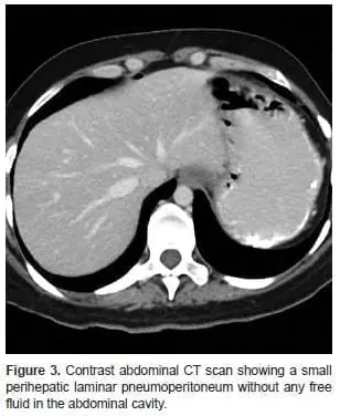
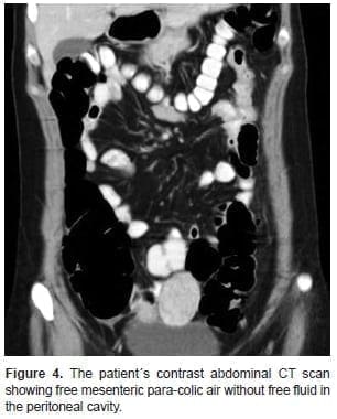
Taking the patient`s stability and absence of peritoneal irritation into account, we chose a conservative approach and started intravenous fluid therapy, analgesics, gastric protection, antibiotics (piperacillin-tazobactam). Abdominal CT showed alteration of mesenteric fat tissue around the distal third of the descending colon and the proximal sigmoid, as well as adjacent air levels indicating pneumoperitoneum around the left hepatic lobe and para-colic mesenteric air (Figures 3 and 4).
[enc_su_row][enc_su_column size=”1/2″ center=”no” class=””] [/enc_su_column] [enc_su_column size=”1/2″ center=”no” class=””]
 [/enc_su_column][/enc_su_row]
During five days of in hospital observation the patient had progressive decrease of CBC with leukocytes of 11.000, stable hemoglobin levels and was given oral nutrition. At day 7, the patient had adequate oral tolerance, intestinal transit as well as significant decrease in abdominal discomfort, and was discharged. Fifteen days following discharge expressed no abdominal pain and had adequate intestinal transit. Pathology report 2 weeks post-resection showed a tubulovillous adenoma with low grade dysplasia. The patient was seen once more at 8 weeks following discharge and no further interventions were necessary.
Discussion
Post-polypectomy syndrome appears when there is transmural burn and serosal inflammation leading to abdominal pain referred by patients on admission. In absence of perforation, surgery is rarely needed. Our patient presented with a micro-perforation and was managed as a perforated diverticulum with pneumoperitoneum.
Risk factors include right-sided polyps (83%) because of thinner mucosa, polyps larger than 2 cm, and arterial hypertension (believed to be due to endothelial dysfunction). However, our patient did not have any of the usual risk factors. The diagnosis of PPS is in general a clinical diagnosis; abdominal tomography may discard or confirm free peritoneal air. Management is determined by the presence of peritoneal irritation, hemodynamic stability and the individual surgeon’s preference. However, in the absence of acute abdomen and hypotension, conservative management should be considered whenever possible, even when perforation is suspected. Most small perforations presenting without acute abdomen usually resolve spontaneously due to omental adherence, thus surgical treatment is not always necessary. (4-9) When conservative treatment is chosen, strict clinical observation is vital. Patients should be given IV antibiotics covering intestinal flora, follow-up CBC and C-reactive protein, IV fluids to prevent dehydration and electrolyte imbalance, analgesics and nothing by mouth for at least 72 hours. A 5-day in-hospital observation is recommended, as was managed our case. Symptoms usually resolve within 5-7 days and outpatient surveillance should be considered. Literature review shows that free peritoneal air on CT is a known predictor of failure of non-surgical treatment; however, other authors have described success cases in certain patients without hemodynamic instability. (5-8) Thus medical management is feasible in stable patients, however when large amounts of free peritoneal air is present, failure rates reach 57-60%. (5-8) In one study conducted by Dharmarajan et al (13-14), 136 patients presented with perforated diverticulitis, and only 5 (3.7%) required emergency surgery on admission, 7 (5%) required emergency surgery for failed conservative treatment; non-operative treatment in those presenting with free air was successful in (92.5%) of patients. Sallinen et al (13-14) reported that in 132 patients with perforated diverticula, 99% were treated successfully without surgery in absence of pericolic abscess and 62% were managed successfully non-operatively in the presence of distant intraperitoneal air. Free fluid in the Douglas fossa was a clear risk factor for failure, thus success rate for patients treated nonoperatively without risk factors was 86%. When stable, patients taken to surgery should be treated with intestinal resection and anastomosis with or without stoma whereas in unstable patients, a Hartmann resection should be chosen. (6-14) With our patient conservative management was chosen because of hemodynamic stability, absence of peritoneal irritation and a general acceptable condition, even though free peritoneal air confirmed “micro-perforation”. Our patient had complete recovery following careful in-hospital observation, IV antibiotics and nothing by mouth; after 5 days a liquid diet was ordered and adequate tolerance and intestinal transit suggested appropriate intestinal lumen healing and the patient was thus discharged. PPS is a rare complication of colonoscopy polyp resections, when accompanied by pneumoperitoneum, patients can be managed conservatively depending on the presence or absence of peritoneal irritation and hemodynamic stability. In patients presenting with peritoneal irritation with frank acute abdomen or hypotension, exploratory surgery is necessary, however, in stable patients without acute abdomen conservative treatment may be considered. Choosing the right management plan requires careful analysis of each patient, but in selected patients conservative treatment may avoid unnecessary surgical intervention.
Interest conflict
None.
References
1. Shin YJ, Kim YH, Lee KH, Lee YJ, Park JH. CT findings of post-polypectomy coagulation syndrome and colonic perforation in patients who underwent colonoscopic polypectomy. Clinical radiology. 2016;71(10):1030-6. doi: 10.1016/j.crad.2016.03.010.
2. Cha JM, Lim KS, Lee SH, Joo YE, Hong SP, Kim TI, Kim HG, Park DI, Kim SE, Yang DH, Shin JE. Clinical outcomes and risk factors of post-polypectomy coagulation syndrome: A multicenter, retrospective, case–control study. Endoscopy. 2013 (03):202-7. doi: 10.1055/s-0032-1326104.
3. Christie JP, Marrazzo J. “Mini-perforation” of the colon—not all postpolypectomy perforations re
quire laparotomy. Diseases of the colon & rectum. 1991;34(2):132-5.
4. Hirasawa K, Sato C, Makazu M, Kaneko H, Kobayashi R, Kokawa A, Maeda S. Coagulation syndrome: delayed perforation after colorectal endoscopic treatments. World journal of gastrointestinal endoscopy. 2015;7(12):1055. doi: 10.4253/wjge.v7.i12.1055.
5. Dib J. Post-Polypectomy Syndrome. The American Journal of Gastroenterology. 2017;112(2):390. doi :10.1038/ ajg.2016.475.
6. Sartelli M, Catena F, Ansaloni L, Coccolini F, Griffiths EA, Abu-Zidan FM, Di Saverio S, Ulrych J, Kluger Y, Ben-Ishay O, Moore FA. WSES Guidelines for the management of acute left sided colonic diverticulitis in the emergency setting. World Journal of Emergency Surgery. 2016;11(1):37. . doi: 10.1186/s13017-016-0095-0.
7. Fantozzi MA. Sindrome Post-polipectomia endoscópica. Rev. argent. coloproctología. 2009;20(1):23-6.
8. Benson BC, Myers JJ, Laczek JT. Postpolypectomy electrocoagulation syndrome: a mimicker of colonic perforation. Case Rep Emerg Med. 2013;2013. doi: 10.1155/2013/687931.
9. Thirumurthi S, Raju GS. Management of polypectomy complications. Gastrointestinal endoscopy clinics of North America. 2015 Apr 30;25(2):335-57.
10. Jehangir A, Bennett KM, Rettew AC, Fadahunsi O, Shaikh B, Donato A. Post-polypectomy electrocoagulation syndrome: a rare cause of acute abdominal pain. Journal of community hospital internal medicine perspectives. J Community Hosp Intern Med Perspect. 2015;5(5):29147. doi: 10.3402/jchimp.v5.29147.
11. Anderloni A, Jovani M, Hassan C, Repici A. Advances, problems, and complications of polypectomy. Clin Exp Gastroenterol. 2014;7:285-96. doi: 10.2147/CEG.S43084.
12. Ma MX, Bourke MJ. Complications of endoscopic polypectomy, endoscopic mucosal resection and endoscopic submucosal dissection in the colon. Best Pract Res Clin Gastroenterol. 2016;30:749-67.
13. Sallinen VJ, Mentula PJ, Leppäniemi AK. Nonoperative management of perforated diverticulitis with extraluminal air is safe and effective in selected patients. Dis Colon Rectum. 2014;57:875-81. doi: 10.1097/ DCR.0000000000000083.
14. Dharmarajan S, Hunt SR, Birnbaum EH, Fleshman JW, Mutch MG. The efficacy of nonoperative management of acute complicated diverticulitis. Diseases of the Colon Rectum. 2011;54:663-71. doi: 10.1007/ DCR.0b013e31820ef759.
