La obstrucción maligna del tracto de salida gástrico o duodenal, primaria o metastásica, es una enfermedad grave encontrada en pacientes con lesiones neoplásicas avanzadas. Sus síntomas incluyen náuseas, vómitos y distensión abdominal con deterioro clínico progresivo del paciente y de su calidad de vida. Tradicionalmente, la paliación ha sido quirúrgica, pero por el carácter invasivo de la intervención y la pobre condición general de estos pacientes, Weaver (1987) y Potts (1990) han encontrado una tasa de morbilidad de 20-30%. La paliación de estas obstrucciones con las EMA ha sido reportada con resultados prometedores. (Castaño, 2004; Lopera, 2001).
Técnica de colocación de las EMA
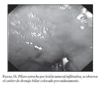
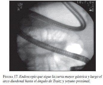
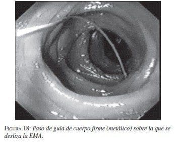
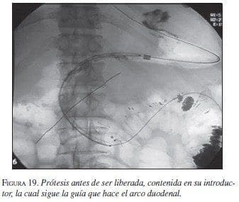
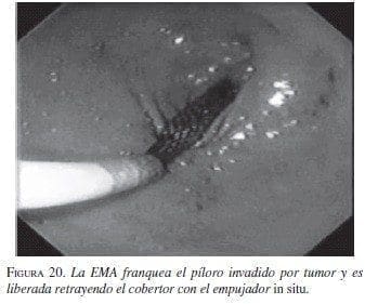
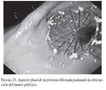
La serie gastroduodenal superior sirve para evaluar la longitud y localización de la estenosis en todos los pacientes. En los casos necesarios se dejó la noche anterior una sonda nasogástrica de descompresión y no se utilizaron antibióticos profilácticos de rutina. El procedimiento se ejecutó bajo sedación consciente, con la cual no se presentaron complicaciones. Con el endoscopio diagnóstico (GIF-P30; Olympus, Tokio) y con anestesia faríngea con xilocaína en aerosol, se visualizó la lesión y cuando era posible se franqueaba con el endoscopio. Se aplicó el material de contraste y el extremo proximal y distal de la estenosis se marcó con infiltración submucosa del contraste o en la piel del paciente con grapas metálicas. Cuando no se pudo franquear la estenosis con el endoscopio, se usó una guía super-stiff de 470 cm (Boston Scientific/Medi-tech) para atravesar la estenosis y dejarla lo más distal posible en el yeyuno. Se seleccionó una prótesis 4 cm más larga que la estenosis para prevenir el crecimiento y obstrucción tumoral en los extremos. Luego de la lubricación con xilocaína, y bajo visión fluoroscópica se colocó a través de la estenosis, el sistema introductor con la prótesis montada. Para la liberación de la EMA, el tubo introductor se retiró mientras el catéter empujador se dejó in situ. Esta maniobra liberó la EMA. La bala plástica se deja dentro del paciente y es expulsada después. No se hizo dilatación con balón ni antes ni después de colocar la prótesis. El seguimiento endoscópico o con fluoroscopia y medio de contraste se hizo para evaluar la permeabilidad y ubicación de la endoprótesis. A los pacientes se les restringió la ingesta a sólo líquidos el primer día, con progresión a dieta líquida completa, blanda o regular según su tolerancia (figuras 16 a 21).
 |
 |
 |
 |
 |
 |
Resultados
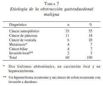
Los pacientes ingresaron al servicio de Gastrohepatología del Hospital Pablo Tobón Uribe y el Hospital San Vicente de Paúl, Universidad de Antioquia entre junio de 1999 y junio de 2004. Se consideraron como inoperables los que mostraban pobre condición física, edad avanzada y extensión regional o a distancia del tumor, o una combinación de estas opciones. Las indicaciones para la colocación de las EMA se presentan en la tabla 7.
Hubo sólo tres fallas técnicas causadas por la longitud inadecuada del sistema introductor o la carencia de una guía de las características descritas en pacientes con estómago marcadamente dilatado que fueron sometidos a gastroyeyunostomías. En cuanto a los síntomas obstructivos previos a la colocación de la EMA, de los nueve pacientes que no toleraban ningún tipo de vía oral, siete mejoraron con la colocación de la misma.
De los dos pacientes que no toleraban la vía oral a pesar de la EMA, El seguimiento radiológico mostró unas prótesis permeables con adecuado paso del medio de contraste a la región distal del duodeno. Estos pacientes estaban en muy mala condición clínica y fueron manejados con alimentación parenteral y medidas paliativas hasta su muerte una semana más tarde. Tres pacientes tenían estenosis muy largas, por lo que requirieron de dos prótesis (una dentro de otra o coaxial).
La migración proximal de la prótesis se observó en tres de las siete totalmente cubiertas, entre uno y cinco días después de su colocación. Estos pacientes presentaron recidiva de sus síntomas obstructivos gastroduodenales. La remoción endoscópica se hizo con un asa de polipectomía de 25 mm (Microvasive Endoscopy; Boston Scientific/medi-tech), seguida por la colocación de una nueva EMA descubierta peroral.
Además, de 50 pacientes con prótesis parcialmente cubiertas, dos presentaron migración. La colonización se observó en dos de las tres EMA descubiertas y en una prótesis parcialmente cubierta dentro de otra en el paciente con mayor seguimiento (96 semanas). Los dos primeros casos con colonización tumoral tenían EMA descubiertas para paliar la obstrucción luego de la migración de las prótesis totalmente cubiertas. El primer paciente tenía una historia de sangrado por el tumor, con oclusión de la EMA por coágulos cuatro semanas después de su colocación. La endoscopia mostró colonización tumoral y el sangrado se manejó con embolización de la arteria gastroduodenal con mejoría. El segundo paciente presentaba síntomas recurrentes de obstrucción gastroduodenal 40 semanas después de la colocación, el estudio baritado de seguimiento reveló estrechez de la EMA por el crecimiento tumoral.
En vista de la condición terminal de ambos pacientes, el manejo fue conservador; ambos toleraron la dieta líquida durante uno y dos meses hasta su muerte. Todos los pacientes fallecieron como consecuencia de sus lesiones tumorales primarias entre 1 y 96 semanas después de la colocación de la EMA (supervivencia promedio de 17,7 semanas).
La sobrevida promedio de los pacientes en quienes no se les pudo colocar la EMA fue de 2,6 semanas, diferencia estadísticamente significativa (p=0,021), comparada con el grupo al que se le colocó la prótesis. El paciente con mayor sobrevida tenía un hipernefroma recurrente, con tres cirugías previas, las dos últimas fallidas; presentó invasión del arco duodenal que se manejó con una prótesis en otra (parcialmente cubiertas) por lo largo de la estenosis, con colonización de la prótesis proximal. Requirió intervención quirúrgica (gastroyeyunostomía), y falleció en el postoperatorio inmediato.
Seguimiento
Al día siguiente del procedimiento a los pacientes se les practicó un esófago-estómago-duodeno, para evaluar la descompresión gástrica y la colocación de la EMA.
Los pacientes fueron controlados a través de llamadas telefónicas semanales y en la consulta externa cada mes, para evaluar la tolerancia a la dieta y la aparición de náuseas, dolor o vómitos. Se practicaron estudios radiológicos con bario o endoscopias en presencia de síntomas sugestivos de recurrencia de la obstrucción. Se determinó el éxito técnico si se colocaba la EMA en el sitio de mayor obliteración por la estenosis. El éxito clínico se definió como la mejoría de los síntomas obstructivos y de la ingesta oral sin necesidad de paliación quirúrgica.
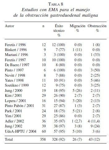
En la tabla 8 se recoge la experiencia publicada con las EMA en el manejo de la obstrucción gastroduodenal maligna.
Self Expandible Metalic Stents in Malignant Esophageal and Gastroduodenal Obstruction
Abstract
Management of malignant obstruction of the gastrointestinal tract constitutes a major challenge. Most patients are in a state of profound depletion due to the underlying malignancy and are not ideal candidates for major operative procedures. In recent years, self-expandible metal stents have shown to be an effective and safe, less invasive alternative in the treatment of malignant esophageal and gastroduodenal obstruction.
We hereby report a prospective study of 99 esophageal and 60 gastroduodenal stents placed over the past four years, together with a literature review.
Technical and clinical success rates were 96% and 85%, respectively. The majority of patients tolerated oral feeding within the first 36 hours following placement of the stent. There were no major complications, such as bleeding or perforation, and there were no deaths. Thirteen patients developed signs of obstruction of the stent, and they were successfully managed by endoscopy. Our data confirm the efficacy of the stent in the palliation of malignant esophageal and gastroduodenal obstruction and the method is associated with low complications rates.
Key words: prosthesis and implants, esophageal neoplasms, stomach neoplasms, endoscopy gastrointestinal.
Referencias
- ACUNAS B, ROZANES I, AKPINAR S, TUNACI A, TUNACI M, ACUNAS G. Palliation of malignant esophageal strictures with self-expanding nitinol stents: drawbacks and complications. Radiology 1996;199:648-52.
- ADLER DG, BARON TH. Endoscopic palliation of malignant gastric outlet obstruction using self-expanding metallic stents: experience in 36 patients. Am J Gastroenterol 2002;97:72-78.
- BETHGE N, SOMMER A, GROSS U. Treatment of esophageal fistulas with a new polyurethane-covered, self-expanding mesh stent: a prospective study. Am J Gastroenterol 1995;90:2143-2146.
- BETHGE N, SOMMER A, VAKIL N, et al. Human tissue responses to metal stents implanted in vivo for the palliation of malignant stenoses. Gastrointest Endosc 1996;43:596-602.
- BETHGE N, SOMMER A, VAKIL N. A prospective trial of self-expanding metal stents in the palliation of malignant esophageal strictures near the upper esophageal sphincter. Gastrointest Endosc 1997; 45: 300-303. Bezzi M, Orsi F, Salvatori FM, Maccioni F, Rossi P. Self-expandable nitinol stent for the management of biliary obstruction: longterm clinical results. J Vasc Interv Radiol 1994;5:287-293.
- BINKERT CA, JOST R, STEINER A, ZOLLIKOFER CL. Benign and malignant stenoses of the stomach and duodenum: treatment with selfexpanding metallic endoprostheses. Radiology 1996; 199:335– CAMPION JP, BOURDELAT D, LAUNOIS B. Surgical treatment of malignant esophagotracheal fistulas. Am J Surg 1983;146:641-6. CASTAÑO R, RUIZ M, SANÍN E. Stent esofágico de nitinol en el manejo de las fístulas esofagorrespiratorias malignas. Revista Col de Gastroenterol 2003, 18 (2): 78-82
- CASTAÑO R, RUIZ M, JULIAO F, SANÍN E, et al. Eficacia de un nuevo stent de nitinol fabricado localmente, en el tratamiento de la obstrucción maligna esofágica. Rev Colom Gastroenterol 2003;18:211-221.
- CASTAÑO R, ÁLVAREZ O, RUIZ M, JULIAO F, SANÍN E, EREBRIE F. Nitinol autoexpandable stent in malignant gastric outlet obstruction. Endoscopy 2004;36 (Suppl I):A242.
- CHAN AC, SHIN FG, LAM YH, et al. A comparison study on physical properties of self-expandable esophageal metal stents. Gastrointest Endosc 1999;49:462-465.
- CHEN JS, LUH SP, LEE F, TSAI CI, LEE JM, LEE YC. Use of esophagectomy to treat recurrent hyperplastic tissue obstruction caused by multiple metallic stent insertion for corrosive stricture. Endoscopy 2000;32:542-545.
- CHOWHAN NM. Injurious effects of radiation on the esophagus. Am J Gastroenterol 1990;85:115-120.
- CONLAN AA, NICOLAOU N, DELIKARIS PG, POOL R. Pessimism concerning palliative bypass procedures for established malignant esophagorespiratory fistulas: a report of 18 patients. Ann Thorac Surg 1984;37:108-110.
- CWIKIEL W, WILLEN R, STRIDHECK H, LILLO-GIL R, VON HOLSTEIN CS. Self expanding stent in the treatment of benign esophageal strictures: experimental study in pigs and presentation of clinical cases. Radiology 1993;187:667-71
- DE BAERE T, HARRY G, DUCREAUX M, et al. Self-expanding metallic stents as palliative treatment of malignant gastroduodenal stenosis. AJR Am J Roentgenol 1997;169:1079-1083.
- DO YS, SONG HY, LEE BH, et al. Esophagorespiratory fistula associated with esophageal cancer: treatment with a Gianturco stent tube. Radiology 1993;187:673-677.
- DURANCEAU A, JAMIESON GG. Malignant tracheoesophageal fistula. Ann Thorac Surg 1984;37:346-354.
- FERETIS C, BENAKIS P, DIMOPOULOS C, et al. Palliation of malignant gastric outlet obstruction with self-expanding metal stents. Endoscopy 1996;28:225-228.
- FERETIS C, BENAKIS P, DIMOPOULOS C, MANOURAS A, TSIMBLOULIS B, APOSTOLIDIS N. Duodenal obstruction caused by pancreatic head carcinoma: palliation with self expandable endoprostheses. Gastrointest Endosc 1997;46:161-165.
- FIORINI AB, GOLDIN E, VALERO JL, et al. Expandable metal coil stent for treatment of broncho-esophageal fistula. Gastrointest Endosc 1995;42:81-83.
- GUKOVSKY-REICHER S, LIN RM, SIAL S, GARRETT B, WU D, LEE T, LEE H, et al. Self-expandable metal stents in palliation of malignant gastrointestinal obstruction: review of the current literature data and 5-year experience at Harbor-UCLA Medical Center. Med Gen Med 2003;10;5:16.
- HAN YM, SONG HY, LEE JM et al., Esophagorespiratory fistula due to esophageal carcinoma: palliation with a covered Gianturco-Stent. Radiology 1996;199:65-79.
- HAUSEGGER KA, KLEINERT R, LAMMER J, et al. Malignant biliary obstruction: histologic findings after treatment with selfexpandable stents. Radiology 1992;185:461-464.
- JEMAL A, MURRAY T, SAMUELS A, GHAFOOR A, WARD E, THUN JM. Cancer Statistics, 2003 CA. 2003;53:5-26.
- JUNG GS, SONG HY, KANG SG, et al. Malignant gastroduodenal obstructions: treatment by means of a covered expandable metallic stent-initial experience. Radiol 2000;216:758-763.
- JUNG GS, SONG HY, SEO TS, PARK SJ, KOO JY, HUH JD, CHO YD. Malignant gastric outlet obstructions: treatment by means of coaxial placement of uncovered and covered expandable nitinol stents. J Vasc Interv Radiol 2002;13:275-283.
- KIM JH, YOO BM, LEE KJ, et al. Self-expanding coil stent with a long delivery system for palliation of unresectable malignant gastric outlet obstruction: a prospective study. Endoscopy 2001;33:838- 842.
- KINSMAN KJ, DE GREGORIO BT, KATON RM, et al. Prior radiation and chemotherapy increase the risk of life-threatening complications after insertion of metallic stents for esophagogastric malignancy. Gastrointest Endosc 1996; 43:196-203
- KOZAREK RA, RALTZ S, BRUGGE WR et al. Prospective multicenter trial of esophageal Z-stent placement for malignant dysphagia and tracheoesophageal fistula. Gastrointest Endosc 1996; 44: 562- 567
- LAASCH HU, NICHOLSON DA, KAY CL, et al. The clinical effectiveness of the Gianturco esophageal stent in malignant esophageal obstruction. Clin Radiol 1998;53:666-672.
- LOPERA J, ALVAREZ O, ZÚÑIGA-CASTAÑEDA A, CASTAÑO R. Initial Experience with Song´s covered duodenal stent in the treatment of malignant gastroduodenal obstruction. J Vasc Interv Radiol 2001;12:1297-1303.
- MACKEN E, GEVERS A, HIELE M, et al. Treatment of esophagorespiratory fistulas with polyurethane-covered selfexpanding metallic mesh stent. Gastrointest Endosc 1996;44:324-326.
- MACCIONI F, ROSSI M, SALVATORI FM, RICCI P, BEZZI M, ROSSI P. Metallic stents in benign biliary strictures: three-year followup. Cardiovasc Intervent Radiol 1992;15:360-366.
- MAETANI I, INOUE H, SATO M, OHASHI S, IGARASHI Y, SAKAI Y. Peroral insertion techniques of self- expanding metal stents for malignant gastric outlet and duodenal stenosis. Gastrointest Endosc 1996;44:468-471.
- MARTINI N, GOODNER JT, D’ANGIO GJ, et al. Tracheo-esophageal fistula due to cancer. J Thorac Cardiovasc Surg 1970;59:319- 324.
- MAYORAL W, FLEISCHER D, SALCEDO J, ROY P, AL-KAWAS F, BENJAMIN S. Nonmalignant obstruction is a common problem with metal stents in the treatment of esophageal cancer. Gastrointest Endosc 2000;51:556-559.
- MAY A, ELL C. Palliative treatment of malignant esophagorespiratory fistulas with gianturco-z stents a prospective clinical trial and review of the literature on covered metal stents. Am J Gastroenterol 1998;93:532-535.
- MCINTEE BE. The wallstent endoprostheses. Gastrointest Endosc Clin N Am 1999;9:373-381.
- MOON T, HONG D, CHUN HJ, JEEN YT, HYUN JH, LEE KB. New approach to radial expansive force measurement of self expandable esophageal metal stents. ASAIO J 2001;47:646-650.
- MORGAN RA, ELLUL JPM, DENTON ERC, GLYNOS M, MASON RC, ADAM A. Malignant esophageal fistulas and perforations: management with plastic-covered metallic endoprostheses. Radiol 1997;204:527-532.
- NELSON DB, AXELRAD AM, FLEISCHER DE, et al. Silicone covered Wallstent prototypes for palliation of malignant esophageal obstruction and digestive respiratory fistulas. Gastrointest Endosc 1997;45:31-37.
- NELSON DB. Expandable metal stents: physical properties and tissue responses. Techn Gastrointest Endosc 2001;3:70-74.
- NEVITT AW, VIDA F, KOZAREK RA, TRAVERSO LW, RALTZ SL. Expandable metallic prostheses for malignant obstructions of gastric outlet and proximal small bowel. Gastrointest Endosc 1998;47:271-276.
- NOMORI H, HORIO H, IMAZU Y, SUEMASU KC. Double stenting for esophageal and tracheobronchial stenoses. Ann Thorac Surg 2000;70:1803-1807.
- PARK HS, DO YS, SUH SW, et al. Upper gastrointestinal tract malignant obstruction: initial results of palliation with a flexible covered stent. Radiology 1999;210:865-870.
- PINTO IT. Malignant gastric and duodenal stenosis: palliation by peroral implantation of a self-expanding metallic stent. Cardiovasc Intervent Radiol 1997; 20:431–434.
- PINTO PABÓN IT, DÍAZ LP, RUIZ DE ADANA JC, LÓPEZ HERRERO J. Gastric and duodenal stents: follow-up and complications. Cardiovasc Intervent Radiol 2001;24:147-153.
- POTTS JR, BROUGHAN TA, HERMANN RE. Palliative operations for pancreatic carcinoma. Am J Surg 1990;159:72-78.
- RAIJMAN I, SIDDIQUI I, AJANI J, LYNCH P. Palliation of malignant dysphagia and fistulae with coated expandable metal stents: experience with 101 patients. Gastrointest Endosc 1998; 48:172- 179.
- SAXON RR, BARTON RE, KATON RM, et al. Treatment of malignant esophagorespiratory fistulas with silicone-covered metallic Z stents . J Vasc Interv Radiol 1995;6:237-242.
- SIERSMA PD, HOP WCJ, DEES J, et al. Coated self-expanding metal stent versus latex prostheses for esophago-gastric cancer with special reference to prior radiation and chemotherapy: a controlled prospective study. Gastrointest Endosc 1998;47:113.
- SILVIS SE, SIEVERT CE, VENNES JA, et al. Comparison of covered versus uncovered wire mesh stents in the canine biliary tract. Gastrointest Endosc 1994;40:17-21.
- SOETIKNO RM, CARR-LOCKE DL. Expandable metal stents for gastricoutlet, duodenal, and small intestinal obstruction. Gastrointest Endosc Clin N Am 1999;9:447-458.
- SONG HY, DO YS, HAN YM, et al. Covered expandable esophageal metallic stent tubes: experiences in 119 patients. Radiology 1994;193:689.
- SUMIYOSHI T, GOTODA T, MURO K, REMBACKEN B, GOTO M, SUMIYOSHI Y, ONO H, SAITO D. Morbidity and mortality after self-expandable metallic stent placement in patients with progressive or recurrent esophageal cancer after chemoradiotherapy. Gastrointest Endosc 2003; 57:882-885.
- VAN DEN BONGARD HJ, BOOT H, BAAS P, TAAL BG. The role of parallel stent insertion in patients with esophagorespiratory fistulas. Gastrointest Endosc 2002;55:110-115.
- VAKIL N, GROSS U, BETHGE N. Human tissue responses to metal stents. Gastrointest Endosc Clin N Am 1999;9:359-365.
- VAKIL N, MORRIS AI, MARCON N, SEGALIN A, et al. A prospective, randomized, controlled trial of covered expandable metal stents in the palliation of malignant esophageal obstruction at the gastroesophageal junction. Am J Gastroenterol 2001;96:1791- 1796.
- WEAVER DW, WIENCEK RG, BOUWMAN DL, WALT AJ. Gastrojejunostomy: is it helpful for patients with pancreatic cancer? Surgery 1987;107:608-612.
- WEIGERT N, NEUHAUS H, RÖSCH T, et al. Treatment of esophagorespiratory fistula with silicone-coated self-expanding metallic stents. Gastrointest Endosc 1995;41:490-496.
- WU WC, KATON RM, UCHIDA T et al., Silicone-covered self-expanding metallic stents for the palliation of malignant esophageal obstruction and esophagorespiratory fistulas: experience in 32 patients and a review of the literature. Gastroint Endosc 1994;40:22-33.
- YATES MR, MORGAN DE, BARON TH. Palliation of malignant gastric and small intestinal strictures with self-expandable metal stents. Endoscopy 1998; 30:266–272.
- YIM HB, JACOBSON BC, SALTZMAN JR, JOHANNES RS, BOUNDS BC, LEE JH, SHIELDS SJ, et al. Clinical outcome of the use of enteral stents for palliation of patients with malignant upper GI obstruction. Gastrointest Endosc 2001;53:329-332.
- YU YT, YANG G, LIU Y, SHEN BZ. Clinical evaluation of radiotherapy for advanced esophageal cancer after metallic stent placement. World J Gastroenterol 2004;10:2145-2146.
Correspondencia:
RODRIGO CASTAÑO LLANO, MD.
rcastanoll@epm.net.co
Medellín, Colombia
